49 Introduction to Memory
Chapter Outline
- How Memory Functions
- Spatial Memory
- Forgetting and Memory Errors
- Memory and the Brain
- Ways to Enhance Memory
Most of us would agree that memory is one of our most valuable assets. Our lives would be empty if we couldn’t store new information and our ability to access past experiences is a big part of who we are. Our nervous systems are bombarded with information that the brain continually sorts, organizes, stores, and retrieves throughout our lifetimes. Unlike physical storage devices, the brain can’t add another filing cabinet or hard drive. Instead, there are many different neural systems that overlap and influence each other to create and retrieve memories, which makes it complicated to study. As a result, scientists combine techniques that allow the study of areas in isolation with techniques that allow the study of memory systems.
One way to study an isolated region is to intentionally damage or remove it, a technique called ablation or lesioning. If the subject shows impaired performance on a memory task after lesioning, then the damaged area was likely involved in that task. Because it would be unethical to create lesions in healthy human subjects, lesion experiments are performed in non-human animals, often to create a model of human systems. This technique allows scientists to assess memory before damage occurs, ensuring that changes in performance are a result of the lesion. Experimental lesions are also specific in location, which isn’t often the case with naturally occurring brain injury. In contrast, studies of brain damage in humans rely on individuals who already have brain damage, or who are having brain regions removed for medical purposes. In these cases, we can’t be sure which effects result from the damage, or which specific regions are responsible.
Still, people with rare brain injuries can provide unique information about the nature of memory systems by examining the abilities that they lost. One famous case study of memory comes from a man named Henry Molaison, known as patient HM, and his collaboration with researcher, Brenda Milner. HM had severe seizures originating in his temporal lobes which were unresponsive to high doses of anticonvulsants, so he underwent an experimental neurosurgery in the care of Dr. William Scoville in 1955. During the surgery, Dr. Scoville removed portions of the medial temporal lobes, specifically, the anterior hippocampus, the parahippocampal gyrus, and the amygdala. Later however, it was discovered that the lesion was smaller than originally thought (Annese et al., 2014). After the surgery, HM’s epilepsy was controlled, but he showed profound and unexpected impairments in memory (Milner, 2005).
HM’s personality was unchanged, and he even scored higher on the Weschler IQ test, likely because his seizures had stopped. He still had early-life memories and retained semantic or fact-based knowledge from the years before the surgery, but he lacked specific, personal memories of that time. Strikingly, he couldn’t remember Dr. Scoville himself, whom he’d known for many years. His most severe impairment however, was an inability to form new, conscious memories.(Milner, 2005; Scoville 1954). At any given moment, HM described his world as being “clear” but said he felt like he was always waking up from a dream (Milner, 2005). After surgery, HM was able to gradually learn some new information with repeated exposure, like the existence of space travel, but this was effortful. For example, it took about five years to navigate the inside of the house he moved to after his operation (Milner, 1970). He was also able to remember small chunks of information if he could continuously repeat it to himself, a process called maintenance rehearsal (Annese et. al, 2014).
Given the unexpected results of HM’s surgery, Scoville invited Brenda Milner, a researcher from the Montreal Neurological Institute with expertise in the function of the medial temporal lobes (Milner, 2005). To test HM’s short term or working memory, Milner adapted a spatial task that asked participants to discover the correct route through a digital maze along a series of “stepping stones”. The participant selects a stone, and if the stone is correct they are given feedback and allowed to continue. If it isn’t correct, they must go back to the start and try again. Most people can remember enough correct steps to make it through the maze after about 20 trials, but HM failed to remember anything after 215 attempts (Milner, 1966; Milner 2005). Milner later trained him on a much shorter version of the maze with only six choice points. Although it took 155 trials and a lot of effort, HM was eventually able to complete three errorless runs through the maze, and even remembered some of the correct pathways two years later (Milner et al., 1968; Milner, 2005).
This digital maze study revealed two key points. First, HM was able to remember the pathway once the stepping stones were reduced to six, so he had some capacity for immediate memory. This indicates that his immediate memory functioned normally even though he couldn’t form longer-term memories. Second, he kept the memories that he was able to create for years, which suggests that forming memories and keeping memories could require different processes. Because HM was missing his medial temporal lobes, we can assume that this area of the brain is important for forming memories, but not necessarily storing them. This case demonstrates a study of memory systems, as HM’s surgery affected multiple areas contributing memory processes. With careful study design, we can make links between brain areas and the behaviours they’re involved in, even if the damaged area is not specific.
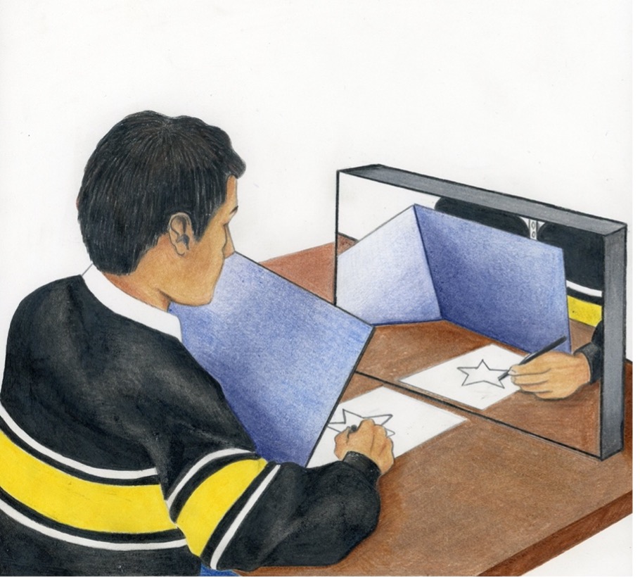
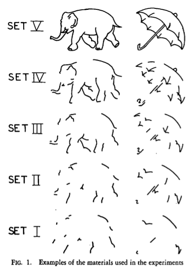
By observing what HM was not able to do, Milner and her team could determine what the medial temporal lobe did control. Milner was also able to determine which parts of memory were not controlled by the medial temporal lobes through her discovery of HM’s capacity for implicit learning, the act of gaining new skills without conscious awareness. First, he completed a motor task where he had to copy a drawing by only looking at a reflection of his hand through a mirror (Figure M.1, above). After 30 trials over three days, he had no conscious awareness of doing the task before, but his performance improved significantly (Corkin 1968; Milner, 1970; Milner, 2005). For the second task, HM had to identify an object shown as multiple incomplete line drawings, each differing in its level of completion (Figure M.3, below). It was nearly impossible for him to identify objects from the most incomplete drawings, but with practice he needed fewer cues to name the items (Gollin, 1960; Milner, 1970; Milner 2005). These studies showed that the medial temporal lobes were not required for these types of memory tasks.
Thanks to Brenda Milner’s work, we’ve learned a lot from HM himself. After dedicating 30 years of his life to the research of memory, Henry Molaison also donated his brain for further study upon his death in 2008 (New York times, 2009), and 2041 high resolution images of his brain sections are now compiled in a virtual brain atlas available to researchers online (Annese et al., 2014) . Despite his contributions to science, it was only after his death that HM became known by his full name, Henry Molaison, rather than his initials. His collaboration with Brenda Milner is now one of the most influential in neuroscience, helping to link the medial temporal lobe with specific memory function and aiding in distinguishing different types of memory.
Link to Learning
To learn more about HM’s life and the research that grew from this case study, visit the Brain Observatory’s Project HM: https://www.thebrainobservatory.org/projecthm
HM’s case provided crucial information in early neuroscience, but we must note that his brain was atypical because of his epilepsy. In fact, post-mortem examination revealed a number of non-surgical lesions that could have contributed to his impairments (Annese et. al, 2014). In the decades following, we have continued to develop techniques for the study of memory. We can now observe memory processes and systems in real time using functional imaging techniques, and even create ‘virtual’ lesions by temporarily interfering with activity in specific brain regions using Transcranial Magnetic Stimulation to study the function of isolated areas (Brem et. al, 2013). Using combinations of these techniques with non-human animal models, as well as other unique cases of brain injury, we can continue to identify the functions of specific areas while also refining our understanding of how they work together.
