9.2 Sensing Hypoxic and Anoxic Conditions
KEY CONCEPTS
By the end of this section, you will be able to do the following:
- Apply your understanding of cell signalling pathways (Chapter 5.6) to explain two examples of signal reception and transduction involved in detecting hypoxic stress.
- Evaluate how the presence of oxygen is related to hydroxylation of HIF-1α.
- Use your understanding from Chapter 9.1 to explain why hypoxia would impede effective electron transport through the mitochondrial ETS.
On a cellular level, changes in oxygen availability stimulate the activity of hypoxia-sensitive genes, which in turn trigger short-term and long-term responses. A cell can sense hypoxic and anoxic conditions through signaling pathways. In animals, there are specific cells dedicated to sensing oxygen deficits. Low oxygen detection pathways are complex and interconnected, with multiple layers of regulation and feedback loops. Malfunctions of these pathways are connected to many human health conditions, such as cancer, cardiovascular disease, and neurological disorders. Thus, understanding the mechanisms by which cells sense and respond to hypoxic/anoxic conditions is an active area of research. Several oxygen sensing pathways in cells are used to detect hypoxic conditions, including hypoxia-inducible factors, prolyl hydroxylase enzyme, and the mitochondrial electron transport system. If you need to review the basics of cell signalling, refer back to Chapter 5.6.
Oxygen Sensing by Hypoxia-Inducible Factors
Hypoxia-Inducible Factors (HIF) are a group of transcription factors that play a central role in the cellular response to hypoxia through the regulation of gene expression. There are three major isoforms (versions) of HIF that are restricted to metazoans (animals), each of which has unique properties and functions: HIF-1, HIF-2 and HIF-3. Major differences between these isoforms lie in their distribution, target genes and regulation by oxygen levels. While HIF-2 and HIF-3 have higher specificity for target genes in different cell types, HIF-1 is ubiquitously expressed and the most studied isoform.
HIF-1 consists of two subunits, HIF-1α and HIF-1β, which heterodimerize by combining two different monomers, to work together and activate the transcription of genes involved in cellular response to hypoxia (Figure 9.4). HIF-1 is only functional when HIF-1α and HIF-1β can bind to each other, which generally does not happen under normoxia. The HIF-1β (aryl hydrocarbon receptor nuclear translocator) subunit is constantly expressed, independent of oxygen availability. However, under normoxic conditions any synthesized HIF-1α is promptly hydroxylated (addition of OH groups) and degraded. Prolyl hydroxylase domain proteins (PHDs) help drive the degradation of HIF-1 α because these enzymes catalyze the hydroxylation of HIF-1α, using oxygen a substrate. This covalent modification of HIF-1α triggers the binding of the von Hippel-Lindau protein (VHL). This marks HIF-1α for degradation by proteasomes (see Chapter 14), preventing its accumulation in the cell.
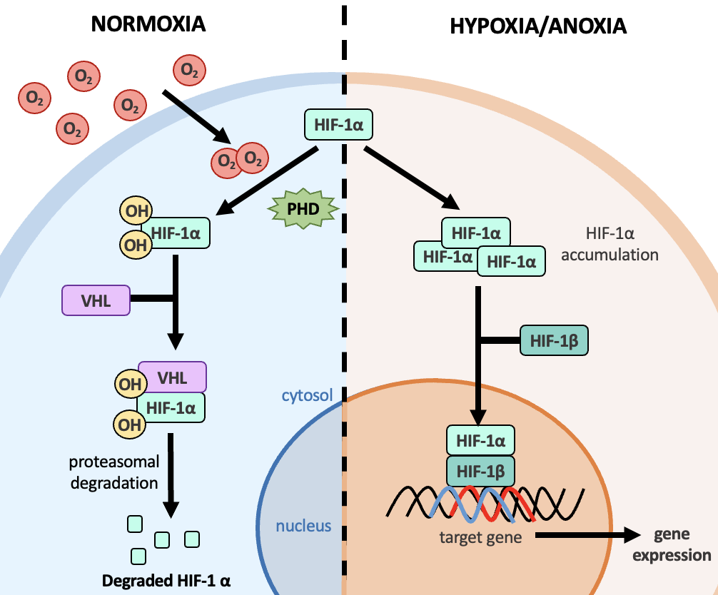
The HIF-1α subunit is accumulated only under hypoxic conditions, when there is limited oxygen available for hydroxylation of HIF-1α by PHDs. The accumulation of HIF-1α allows it to bind with HIF-1β, making the HIF-1 transcription factor functional and active. In the nucleus, the HIF-1αβ complex binds to specific DNA sequences, known as hypoxia-response elements (HRE), found in the promoter region of target genes (Figure 9.4). This leads to the upregulation of gene expression which encode proteins involved in a wide range of processes to support hypoxia tolerance.
Link to Learning
Oxygen Sensing by the Electron Transport System
As previously discussed, the mitochondrial electron transport system (ETS) is crucial to cellular respiration and energy production in the cell. The ETS uses oxygen as a substrate and therefore plays a role in sensing hypoxic conditions. Under decreased availability of oxygen, electron transport through the ETS becomes less efficient, resulting in increased production of reactive oxygen species (ROS) by the ETS (for more about ROS, consult Chapter 10). It is hypothesized that, as a result, this ROS production signals the cell of low oxygen conditions via multiple pathways such as HIF-1. Thus, while the ETS pathway does not directly detect the molecular oxygen levels, it is indirectly involved in hypoxic detection by altering the production of ROS.
